Lesions seen in Chicken Infectious Anemia
Lesions:
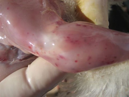
- Focal lesions (mostly in the wings) appear as ecchymotic skin haemorrhages.
- The lesions turn blue and may break, releasing serosanguineous exudate which is prone to secondary bacterial infections, leading to gangrenous dermatitis. This can be especially notorious at the end of the wings, hence the name “Blue Wing Disease” used to describe this condition. The tips of the wings may appear haemorrhagic and necrotic.
- The mortality peaks 5 -6 days after the appearance of the clinical signs, declining to normal 5 – 6 days later.
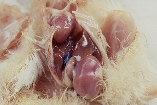
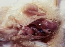
- Thymus atrophy with lobes appearing small and greyish. When observed closely, the medullar part of the lobes predominate over the cortical part.
- Bone marrow atrophy. When observed closely it appears pale.
Histology
Bone marrow
In the bone marrow a decrease in the number haematopoietic cells is observed 4 – 6 days post infection. This is followed by the appearance of large blastic cells. The haematopoietic tissue is replaced by adipose tissue; this gives the bone marrow a pale appearance.
Other organs
Depletion of lymphocytes from the thymus, spleen, Bursa Fabricius and caecal tonsils is observed, followed by hyperplasia of reticular cells. Changes in the thymus appear at 4 days post infection, with cortical lymphocytes disappearing and being replaced by reticular cells. Medullar lymphocytes are also reduced. Recovery starts 20 days post infection in convalescent birds. The changes found in the different organs suggest that CAV multiplies in the lymphocytes.
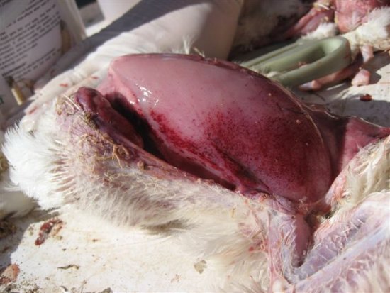
Pale carcass
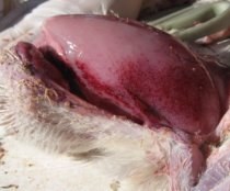
Subcutaneous haemorrhages
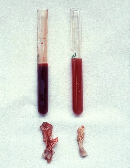
Pale bone marrow and watery blood